
Brain symmetries and gauge theories
Arturo Tozzi, MD, PhD
tozziarturo@libero.it
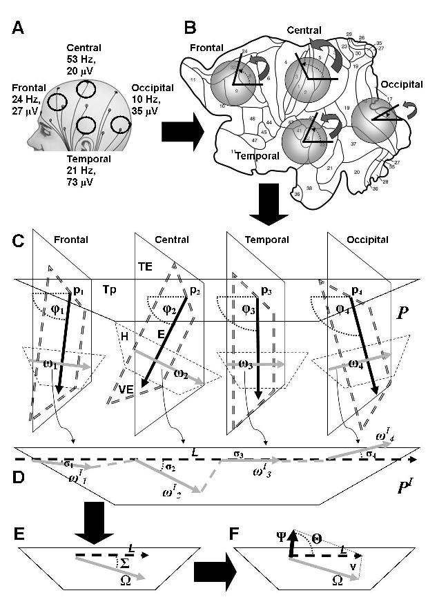
Figure. See text for details.
INTRODUCTION
Two focuses highlighting current technical advances in neuroscience have been recently published (Dayan et al., 2013; Mattei, 2014). The exciting innovations call for multidisciplinary theoretical approaches, such as the assessment of highly organized biological model through physical/mathematical procedures. The aim of our theoretical paper is therefore to examine the possibility that part of brain electric activity is driven by one or more continuous forces, called the “gauge fields”, coming from outside the cortex.
A gauge field in physics is embodied in a “gauge theory”. Gauge theories state that some features of a system are invariant under a continuous group of “local” transformations (Zeidler, 2011). The cornerstone of gauge theories are the continuous symmetries, expressed in physics by a function that summarizes the dynamics of the system, called the Lagrangian.
Are we allowed to sketch a gauge theory of brain function? The answer is yes, because cerebral electric activity displays a feature typical of a gauge theory: a symmetry, and specifically a “continuous scale” symmetry. Indeed, an high number of brain currents’ possible source configurations in different cortical areas (“local transformations”, such as gamma oscillations in somatomotor cortex during states of enhanced vigilance, or alpha waves in posterior zones with eyes open) gives rise to a set of measured potentials characterized by a general scaling property. It has been demonstrated that cerebral activity's frequency spectrum exhibits a scale-invariant behavior S(f)= 1/fn, where the power spectrum is S(f), the frequency is f and the power spectral density is n (which values equals the slope of the line times -1 in a log power versus log frequency scatter plot) (Milstein et al., 2009). The 1/fn fractal like structure is a parameter which summarizes the values of the frequency and amplitude of cerebral areas in a given time and gives rise to such complex scale-free system. We will try to explain this structure via the superimposition of a gauge field. We indeed hypothesize that the 1/fn fractal like structure is not an intrinsic feature of neuronal activity, but that it is regulated by a force external to the system.
In an hypothetical model of gauge theory of brain function, the global invariant 1/fn scaling stands for the complete Lagrangian density L of the total brain currents and is therefore regarded as a global symmetry, while the local changes in frequency and amplitude are regarded as local gauge symmetries. In gauge theories, the global symmetry of the system is maintained, although the presence of local changes of symmetry, by a force acting on the system from outside, called the gauge field. Some data in literature point towards the influence of external forces on brain currents: e. g. a recent paper suggests that neural events locked to a basic physiological input such as heartbeats may underlie behaviorally relevant differential activation in multifunctional cortical areas (Park, 2014).
In our theoretical paper, we will discuss how this hypothetical gauge field could be detected and measured. Two steps are needed to achieve this: first, neurophysiological procedures; then, differential geometry.
A FEASIBLE THEORETICAL EXPERIMENTAL APPROACH
In order to quantitatively assess the required gauge fields, we propose to carry out simple neurophysiologic procedures in humans. The use of a non invasive electroencephalogram (EEG) could be acceptable, because it exhibits significant 1/f-like power spectrum scaling (Pritchard, 1992). Subjects undergo 10-20 EEG measurements during active concentration and rest, eyes open and closed. In Figure 1A, just as an example, we chose four random cerebral areas with hypothetical EEG values of frequency and amplitude. The n value of recorded EEG segments could be estimated by fractal analyses (Ihlen 2012). The most interesting cognitive phenomena occur in time windows shorter than seconds (Buzsáki and Watson, 2012); consequently, EEG frequencies and amplitudes need to be evaluated in a chosen time of subseconds.
Once achieved the data from each point of the cortex, the EEG electric brain activity is investigated in the language of differential geometry: in summary, we will describe the dynamics of cortical electric fields in terms of sections of fiber bundles, jet manifolds and Ehresmann connections. The procedure is the following. Cerebral hemispheres are unfolded and flattened into a two-dimensional reconstruction by computerized procedures (Van Essen, 2005), allowing the entire cortical surface to be transferred to an atlas M of CK (differentiable), C∞ (smooth), finite dimensional manifolds, each one mapping a brain area (Figure 1B). The set of power spectra describing each cortical area’s frequencies and amplitudes stands for a continuous group of local transformations acting on sections of M. M is thus equipped with a constant matrix G belonging to the SO(3) Lie group, isomorphic to the rotation group of the sphere (Figure 1B).
M is a principal G-bundle P characterized by a trivial, smooth and differentiable fiber bundle, by vector bundles E and by a tangent bundle TE (Figure 1C). The electrical forces are described by numbers, arranged in vectors and angles, representing the action G on a chosen local section E of P. Four forces G are depicted in the concise Figure 1C as four vector bundles E arising from four points p in the tangent space Tp. They are equipped with four n-dimensional rotation angles φ (φ1, φ2, …,φn)T standing for the local 1/fn scaling of each of the four brain areas. Rotations through tiny angles link nearby transformations of angles φ arising from points p: as a result, the linear approximation of the function G at p (and its angle φ) in each dimension can be described by introducing a partial derivative. In brief, changes in degrees of φ in selected brain areas match with different power spectra and hence with different electric configurations.
In a gauge theory, the geometric “link” between L and φ can be defined in terms of a connection form, the Ehresmann connection (Ehresmann, 1950). If we identify the horizontal space H, perpendicular to the vertical space VE, we can extrapolate the Ehresmann connection ω, which is a vector on TE (Figure 1C). The Lagrangian density L is indeed a function of TE and H. It would be correct to formulate all rates of change of ω and φ in terms of covariant derivative, a linear differential operator in each associated TE which allows different points (and their angles) to be compared.
Mapping by affine connection every vector ω of P into the bijective, diffeomorphic PI space, a curvature form is constructed (Figure 1D). When the vectors ωI intersect the unique horizontal lift, which corresponds to the invariant L, the angles σ are obtained.
The behavior of the vectors ωI and angles σ can be compactly written by pointwise vector addition of the partial derivatives of the function G at each point. As a result, we get a single vector: + … . The angle Σ is introduced (Figure 1E), which stands for the interaction Lagrangian Lint and expresses the 1/fn scaling values of vector addition obtained by our experimental procedure. If the lines L and Ω are parallel, Σ equals the zero, Lint equals L and the symmetry of the system is preserved.
Otherwise, if L and Ω are not parallel (it happens nearly always in phisiological systems), Σ is different from zero, Lint is different from L and the system displays a broken or absent symmetry. In this case, in order to ensure the invariance of L and to restore the symmetry, we need to define a covariant derivative such that the derivative of Σ will again transform identically with Σ. According to the covariant version of gauge theories, the correction terms are reinterpreted as couplings to an additional divergent counter term, the gauge field, by allowing the symmetry parameter to vary from place to place in the local coordinate system. Figure 1F shows the procedure in a very abridged form. If we do not to take into account L and look at the sole vector Ω and its angle Σ, we observe nothing else than a single force. If we instead regard L as a vector which basis results from the scalar components of its vector space v, Ω (and its angle Σ) turns out to be just one of the covariant components of L. In order to keep L invariant, we need to add another component into v: hence we introduce the vector Ψ, equipped with the angle Θ (Figure 1F). The angle Θ stands for the gauge field Lagrangian Lgf and expresses the global value of the required gauge field’s 1/fn scaling. We are then allowed to make accurate predictions of forces: we may extrapolate from Θ the values of time, frequency and amplitude of the gauge field required to keep invariant the 1/fn scaling during cortical activity.
CONCLUSIONS
The above described procedure allows us to achieve the values of the hypotetical gauge field acting on brain currents. The further step will be to investigate where gauge field comes from (from the human body or from the surrounding environment). We speculate that it could be situated either in structures internal to the body but outside the cortex (such as the heart rate, or the ascending arousal system) or in the external environment (sensory afferences).
A deeper knowledge of gauge symmetries will lead to novel approach to organized biological models, will improve our understanding of brain functions and will pave the way to innovative therapeutic strategies. A feasible field of application is epilepsy: seizures could be counteracted by appropriate artificial gauge fields (e.g., transcranial direct current stimulation or drugs) able to “recovery” the Lagrangian and restore the electric symmetry.
REFERENCES
1. Buzsáki, G., Watson, B.O. (2012). Brain rhythms and neural syntax: implications for efficient coding of cognitive content and neuropsychiatric disease. Dialogues Clin. Neurosci. 4, 345-367.
- Dayan, E., Censor N., Buch, E.R., Sandrini, M., and Cohen, L.G. (2013). Noninvasive brain stimulation: from physiology to network dynamics and back. Nat. Neurosci. 16, 838–844. doi: 10.1038/nn.3422
- Ehresmann, C. (1950). Les connexions infinit´esimales dans un espace fibrée differentiable. Colloque de Topologie, Bruxelles, pp. 29–55.
4. Ihlen, E.A. (2012). Introduction to multifractal detrended fluctuation analysis in matlab. Front. Physiol. 141, 1-18. doi: 10.3389/fphys.2012.00141
5. Mattei, T.A. (2014). Unveiling complexity: non-linear and fractal analysis in neuroscience and cognitive psychology. Front. Comput. Neurosci. 21; 8:17. doi: 10.3389/fncom.2014.00017
6. Milstein, J., Mormann, F., Fried, I., and Koch, C. (2009). Neuronal shot noise and Brownian 1/f2 behavior in the local field potential. PLoS One. 4, e4338. doi: 10.1371/journal.pone.0004338
7. Park, H.D., Correia, S., Ducorps, A., Tallon-Baudry, C. (2014). Spontaneous fluctuations in neural responses to heartbeats predict visual detection. Nat. Neurosci. 2014 Mar 9. [Epub ahead of print]. doi: 10.1038/nn.3671
- Pritchard, W.S. (1992). The brain in fractal time: 1/f-like power spectrum scaling of the human electroencephalogram. Int. J. Neurosci. 66, 119-129.
- Van Essen, D.C. (2005). A Population-Average, Landmark- and Surface-based (PALS) atlas of human cerebral cortex. Neuroimage. 28, 635–666.
10. Zeidler, E. (2011). Quantum Field Theory III: Gauge Theory. Springer. doi: 10.1007/978-3-642-22421